Imaging the human body's morphological and angiographic information is essential for diagnosing, monitoring, and treating medical conditions...
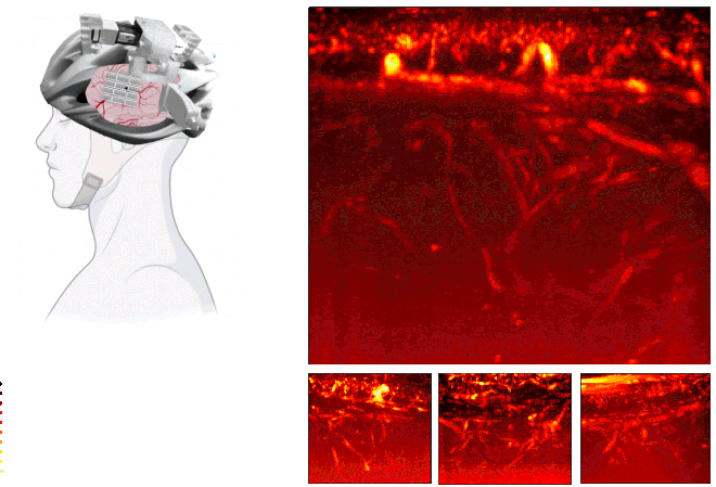
Noninvasive imaging deep into the adult brain at submillimeter and millisecond scales remains a challenge in medical imaging. Here, we present a helmet-based ultrasound system designed for patients post-hemicraniectomy, enabling high-resolution functional brain monitoring...
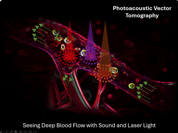
Imaging deep haemodynamics non-invasively remains a quest. Although optical imaging techniques can be used to measure blood flow...
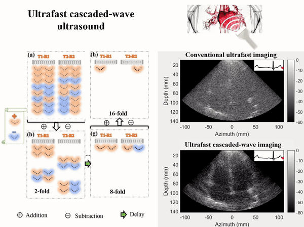
Ultrafast ultrasound imaging using plane or diverging waves, instead of focused beams, has advanced greatly the development of novel ultrasound imaging methods for evaluating tissue functions beyond anatomical information...